Home

Mardi silence nager cytochrome c fluorescence la grève Aidezmoi à quelle fréquence

G/GO induces cytochrome c release from mitochondria in H9c2 cells.... |  Download Scientific Diagram
G/GO induces cytochrome c release from mitochondria in H9c2 cells.... | Download Scientific Diagram

Pharmaceutics | Free Full-Text | Quantifying Cytosolic Cytochrome c  Concentration Using Carbon Quantum Dots as a Powerful Method for Apoptosis  Detection
Pharmaceutics | Free Full-Text | Quantifying Cytosolic Cytochrome c Concentration Using Carbon Quantum Dots as a Powerful Method for Apoptosis Detection
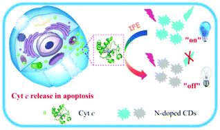
Label-free fluorescence imaging of cytochrome c in living systems and  anti-cancer drug screening with nitrogen doped carbon quantum dots -  Nanoscale (RSC Publishing)
Label-free fluorescence imaging of cytochrome c in living systems and anti-cancer drug screening with nitrogen doped carbon quantum dots - Nanoscale (RSC Publishing)

Immunofluorescence detection of cytochrome c ( A, green fluorescence ),...  | Download Scientific Diagram
Immunofluorescence detection of cytochrome c ( A, green fluorescence ),... | Download Scientific Diagram
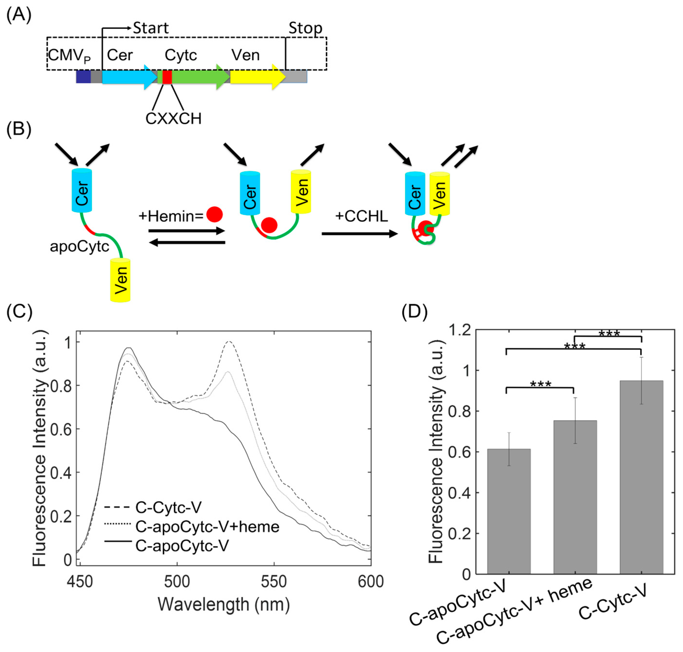
Biosensors | Free Full-Text | Genetically Encoded Fluorescent Probe for  Detection of Heme-Induced Conformational Changes in Cytochrome c
Biosensors | Free Full-Text | Genetically Encoded Fluorescent Probe for Detection of Heme-Induced Conformational Changes in Cytochrome c

Interaction of polyethylene glycol with cytochrome c investigated via in  vitro and in silico approaches | Scientific Reports
Interaction of polyethylene glycol with cytochrome c investigated via in vitro and in silico approaches | Scientific Reports

A) UV−vis spectrum of Cyt c (a) and fluorescence emission spectrum of... |  Download Scientific Diagram
A) UV−vis spectrum of Cyt c (a) and fluorescence emission spectrum of... | Download Scientific Diagram

Cytochrome c release visualized by confocal immunofluorescence... |  Download Scientific Diagram
Cytochrome c release visualized by confocal immunofluorescence... | Download Scientific Diagram

A new quantitative assay for cytochrome c release in apoptotic cells | Cell  Death & Differentiation
A new quantitative assay for cytochrome c release in apoptotic cells | Cell Death & Differentiation

Fluorescence emission of cytochrome-c subjected to different... | Download  Scientific Diagram
Fluorescence emission of cytochrome-c subjected to different... | Download Scientific Diagram

Nitrogen and fluorine co-doped green fluorescence carbon dots as a  label-free probe for determination of cytochrome c in serum and temperature  sensing - ScienceDirect
Nitrogen and fluorine co-doped green fluorescence carbon dots as a label-free probe for determination of cytochrome c in serum and temperature sensing - ScienceDirect

Mitochondrial Apoptosis Assay Cell Line (Green Fluorescent Cytochrome c) –  Cells Online
Mitochondrial Apoptosis Assay Cell Line (Green Fluorescent Cytochrome c) – Cells Online

Emission spectra of (a) BSA (b) Cytochrome-C (c) Ovalbumin (d) Lysozyme...  | Download Scientific Diagram
Emission spectra of (a) BSA (b) Cytochrome-C (c) Ovalbumin (d) Lysozyme... | Download Scientific Diagram

Quenching of TryptophanFluorescence in Unfolded Cytochrome c: A Biophysics  Experiment for Physical Chemistry Students. - Abstract - Europe PMC
Quenching of TryptophanFluorescence in Unfolded Cytochrome c: A Biophysics Experiment for Physical Chemistry Students. - Abstract - Europe PMC
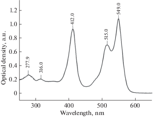
Study of Interaction of Fluorescent Cytochrome C with Liposomes,  Mitochondria, and Mitoplasts by Fluorescence Correlation Spectroscopy |  Russian Journal of Bioorganic Chemistry
Study of Interaction of Fluorescent Cytochrome C with Liposomes, Mitochondria, and Mitoplasts by Fluorescence Correlation Spectroscopy | Russian Journal of Bioorganic Chemistry
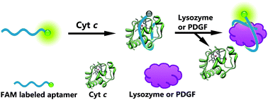
Fluorescence turn-on detection of a protein using cytochrome c as a  quencher - Chemical Communications (RSC Publishing)
Fluorescence turn-on detection of a protein using cytochrome c as a quencher - Chemical Communications (RSC Publishing)
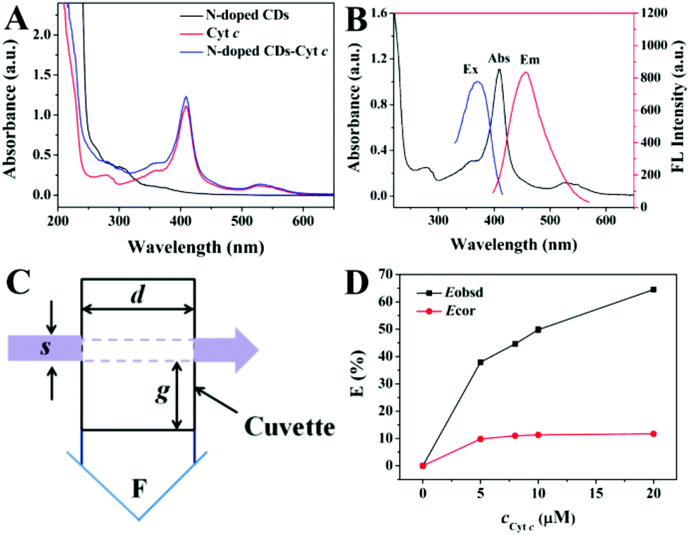
Label-free fluorescence imaging of cytochrome c in living systems and  anti-cancer drug screening with nitrogen doped carbon quantum dots -  Nanoscale (RSC Publishing) DOI:10.1039/C7NR08987B
Label-free fluorescence imaging of cytochrome c in living systems and anti-cancer drug screening with nitrogen doped carbon quantum dots - Nanoscale (RSC Publishing) DOI:10.1039/C7NR08987B
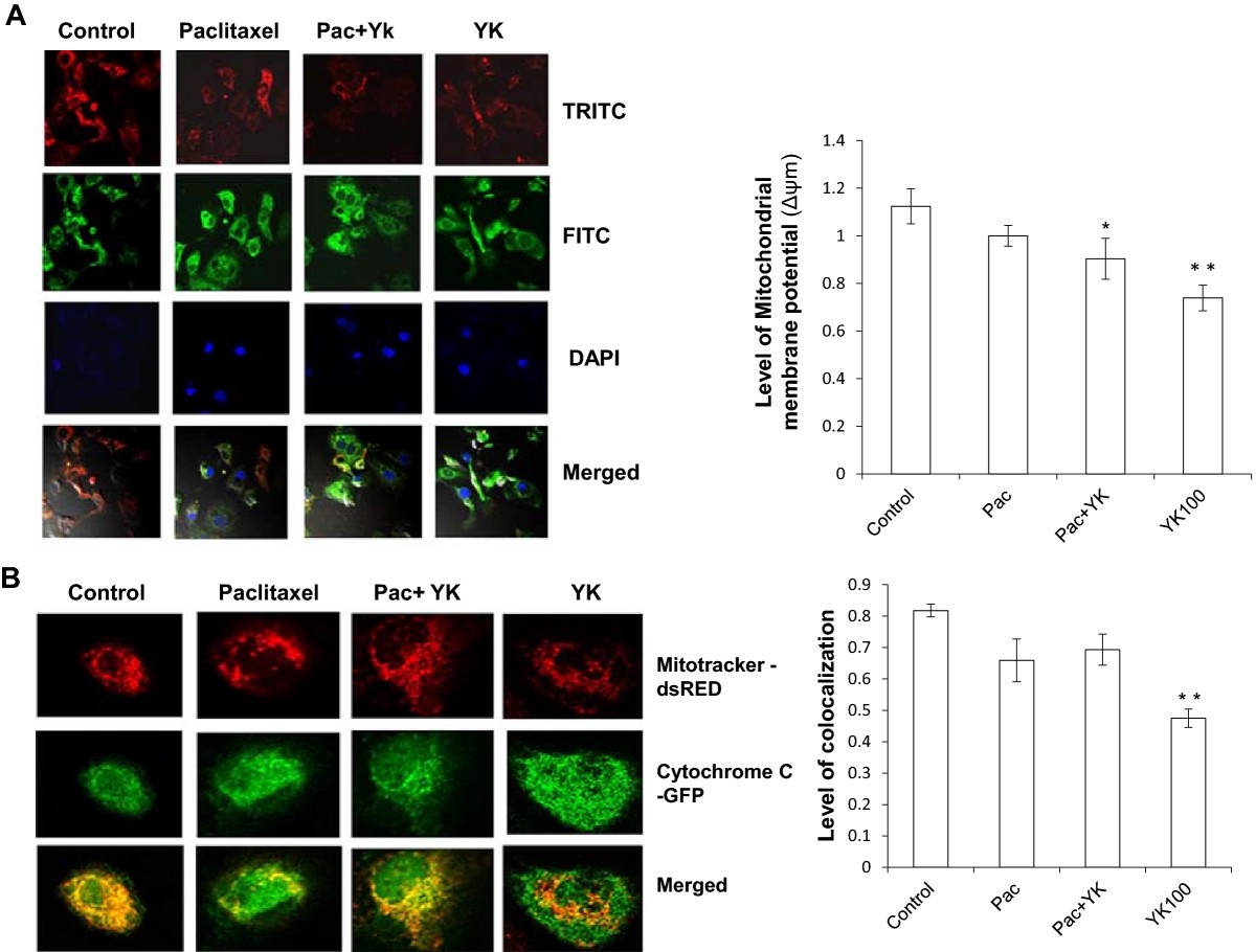
RETRACTED ARTICLE: Molecular insights into the anti-cancer properties of  Traditional Tibetan medicine Yukyung Karne | BMC Complementary Medicine and  Therapies | Full Text
RETRACTED ARTICLE: Molecular insights into the anti-cancer properties of Traditional Tibetan medicine Yukyung Karne | BMC Complementary Medicine and Therapies | Full Text

Fluorescence profiles of a pure suspension of cytochrome c, and of a... |  Download Scientific Diagram
Fluorescence profiles of a pure suspension of cytochrome c, and of a... | Download Scientific Diagram

Figure 3 from Fluorescence activation imaging of cytochrome c released from  mitochondria using aptameric nanosensor. | Semantic Scholar
Figure 3 from Fluorescence activation imaging of cytochrome c released from mitochondria using aptameric nanosensor. | Semantic Scholar

Cells | Free Full-Text | Cytochrome c Oxidase Inhibition by ATP Decreases  Mitochondrial ROS Production
Cells | Free Full-Text | Cytochrome c Oxidase Inhibition by ATP Decreases Mitochondrial ROS Production

Mitochondrial Apoptosis Assay Cell Line (Green Fluorescent Cytochrome c) –  Cells Online
Mitochondrial Apoptosis Assay Cell Line (Green Fluorescent Cytochrome c) – Cells Online

The release of cytochrome c and activation of caspase 3 is delayed in... |  Download Scientific Diagram
The release of cytochrome c and activation of caspase 3 is delayed in... | Download Scientific Diagram
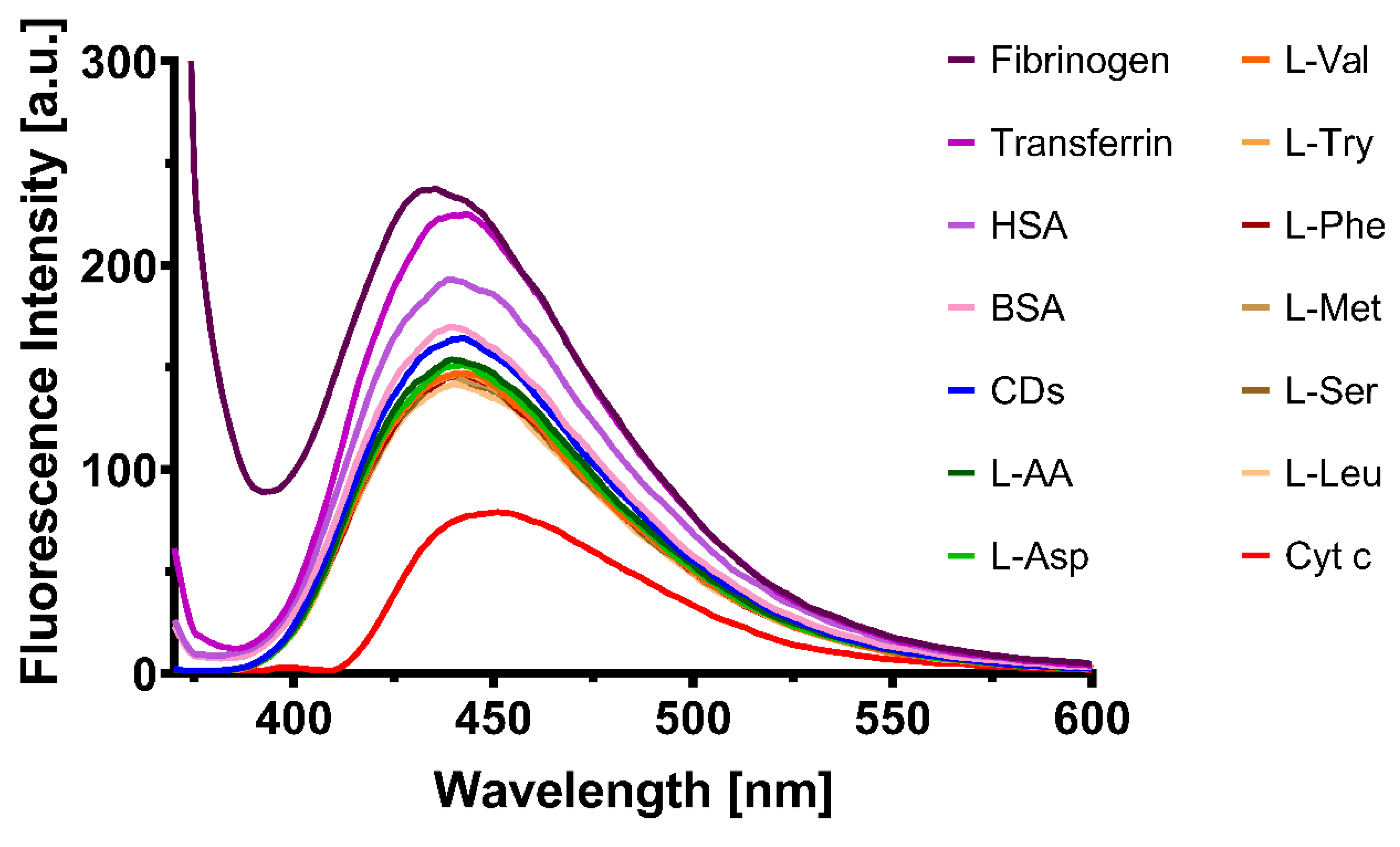
Pharmaceutics | Free Full-Text | Quantifying Cytosolic Cytochrome c  Concentration Using Carbon Quantum Dots as a Powerful Method for Apoptosis  Detection
Pharmaceutics | Free Full-Text | Quantifying Cytosolic Cytochrome c Concentration Using Carbon Quantum Dots as a Powerful Method for Apoptosis Detection