Home

Devenir Clip papillon devise fluorescent nissl stain avaler Taudis Douceur

Neural somata from mouse brain section. NeuroTrace 500/525 green-fluorescent  Nissl stain, DAPI and CellTracker CM-DiI. | Thermo Fisher Scientific - NZ
Neural somata from mouse brain section. NeuroTrace 500/525 green-fluorescent Nissl stain, DAPI and CellTracker CM-DiI. | Thermo Fisher Scientific - NZ

NisslFluor™ Red, Ready-to-Dilute™, Fluorescent Nissl Stain for Healthy  Neurons
NisslFluor™ Red, Ready-to-Dilute™, Fluorescent Nissl Stain for Healthy Neurons

What is a Nissl stain? — Brain Stuff

Propidium iodide (PI) stains Nissl bodies and may serve as a quick marker  for total neuronal cell count - ScienceDirect
Propidium iodide (PI) stains Nissl bodies and may serve as a quick marker for total neuronal cell count - ScienceDirect

NeuroScience Associates | Thionine (Nissl) Cell Body Stain

Pictures
Pictures

Deep-Red Fluorescent Nissl Stain

A fluoro-Nissl dye identifies pericytes as distinct vascular mural cells  during in vivo brain imaging | Nature Neuroscience
A fluoro-Nissl dye identifies pericytes as distinct vascular mural cells during in vivo brain imaging | Nature Neuroscience
NeuroTrace™ 640/660 Deep-Red Fluorescent Nissl Stain - Solution in DMSO

Deep-Red Fluorescent Nissl Stain

NeuroTrace™ 530/615 Red Fluorescent Nissl Stain - Solution in DMSO

CIC | Methodology
CIC | Methodology

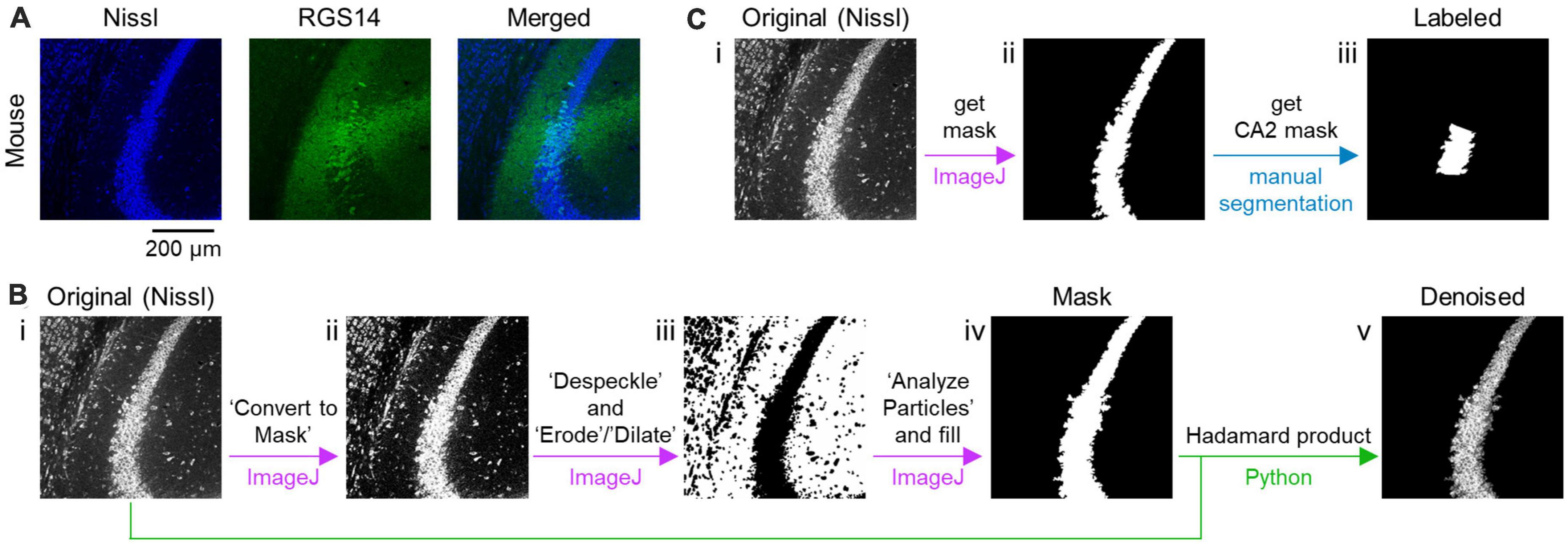
Frontiers | Machine learning-based segmentation of the rodent hippocampal  CA2 area from Nissl-stained sections
Frontiers | Machine learning-based segmentation of the rodent hippocampal CA2 area from Nissl-stained sections

NisslFluor™ Silver, Ready-to-Dilute™, Fluorescent Nissl Stain for Healthy  Neurons
NisslFluor™ Silver, Ready-to-Dilute™, Fluorescent Nissl Stain for Healthy Neurons

Red fluorescent Nissl body imaging using the DUV microscope with (a-c)... |  Download Scientific Diagram
Red fluorescent Nissl body imaging using the DUV microscope with (a-c)... | Download Scientific Diagram

Detects Nissl Expression in Mouse DRG Neurons | Biocompare Antibody Review

Fluorescent Nissl staining of DCN and major cell types at selected... |  Download Scientific Diagram
Fluorescent Nissl staining of DCN and major cell types at selected... | Download Scientific Diagram

CIC | Methodology
CIC | Methodology

Hito Mini NisslStain Kit | Hitobiotec Corp.

The use of Nissl staining for the identification and selection of... |  Download Scientific Diagram
The use of Nissl staining for the identification and selection of... | Download Scientific Diagram

NisslFluor™ Orange, Ready-to-Dilute™, Fluorescent Nissl Stain for Healthy  Neurons
NisslFluor™ Orange, Ready-to-Dilute™, Fluorescent Nissl Stain for Healthy Neurons

Invitrogen™ NeuroTrace™ 500/525 Green Fluorescent Nissl Stain - Solution in  DMSO
Invitrogen™ NeuroTrace™ 500/525 Green Fluorescent Nissl Stain - Solution in DMSO

Nissl Stain - FD Neuro Technologies, Inc.

Fluorescent Nissl staining of the brainstem in P21 mouse. A... | Download  Scientific Diagram
Fluorescent Nissl staining of the brainstem in P21 mouse. A... | Download Scientific Diagram